Robotic repair for selected kidney drainage obstruction, including pelvi-ureteric junction obstruction, planned after imaging review and specialist assessment.
Short-notice appointments may be available
For urgent or last-minute appointment enquiries, please call the relevant secretary team so availability can be checked quickly. Timing is subject to hospital and clinical availability.
Robotic repair for kidney drainage obstruction
Robotic pyeloplasty is an operation to repair a narrowed drainage point between the kidney and ureter. It may be recommended for selected patients with pelvi-ureteric junction obstruction, hydronephrosis, pain, recurrent infection or impaired drainage.
Condition
PUJ obstruction
A narrowed drainage point can cause kidney swelling, pain, infection or reduced drainage.
Assessment
Scan review
Ultrasound, CT, nuclear medicine scans or prior imaging may be reviewed to assess drainage and kidney function.
Repair
Robotic reconstruction
The narrowed segment is repaired to help urine drain more freely where surgery is suitable.
Who this may be suitable for
Selected patients with pelvi-ureteric junction obstruction.
Patients with kidney swelling, recurrent pain, infection or drainage problems.
Cases where investigation suggests a reconstructive repair is appropriate.
What to expect
Review of symptoms, medical history and kidney imaging.
Discussion of robotic repair, alternatives, risks and expected recovery.
Follow-up planning, which may include stent care and repeat imaging where appropriate.
Consultant-led treatment planning
Mr Shahzad explains the likely cause of obstruction, whether robotic repair is suitable and what the expected recovery and follow-up pathway may involve.
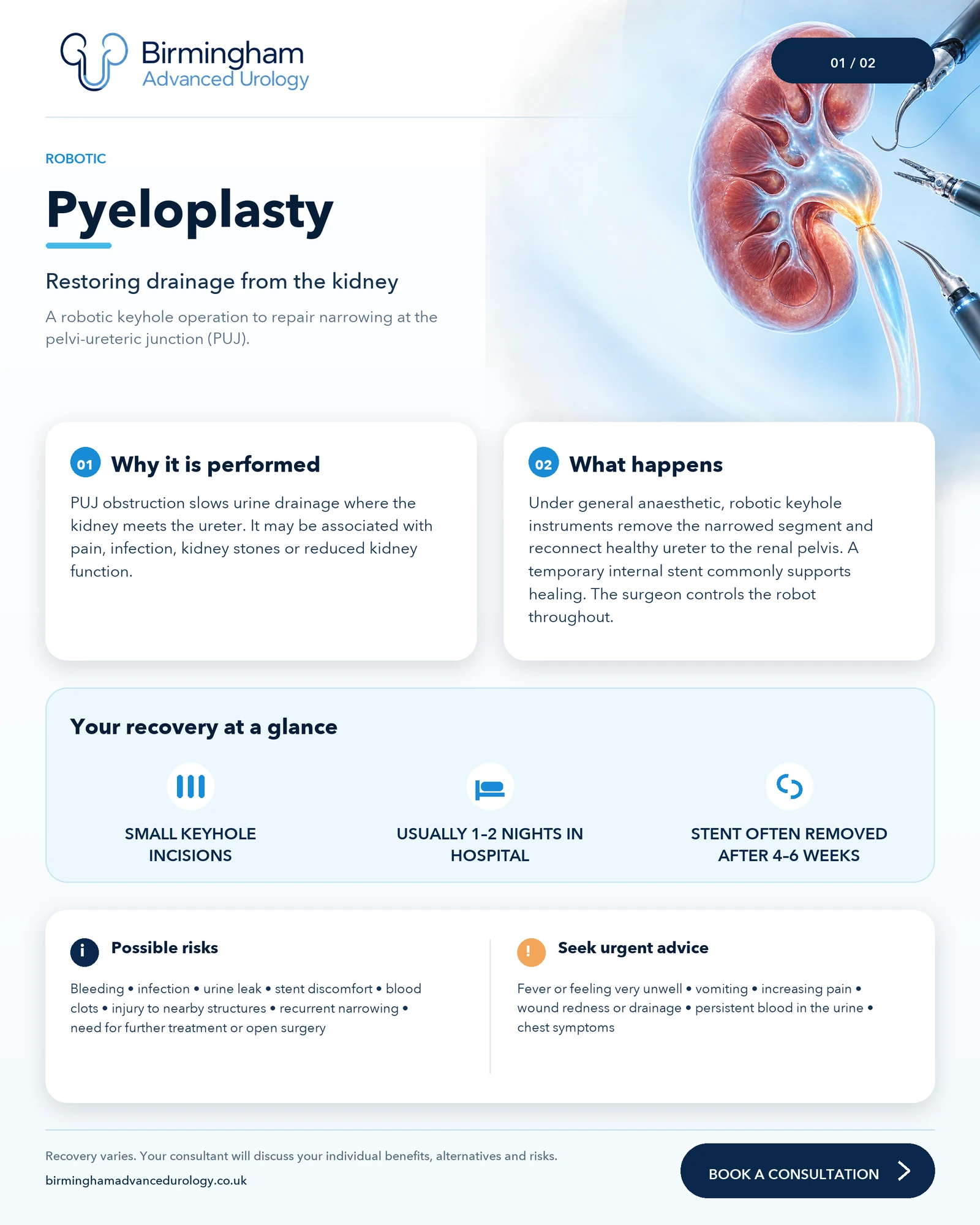
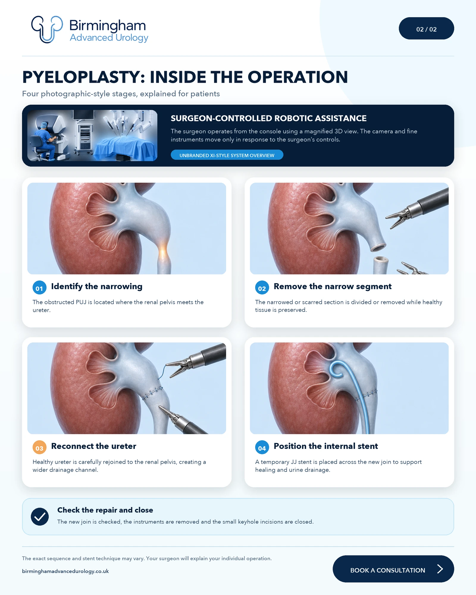
Robotic pyeloplasty: patient visual guide
These patient-friendly illustrations explain why robotic pyeloplasty is performed, how the surgeon controls the robotic system and the main stages of reconstruction. Select either image to view it at full size.
Patient overview: PUJ obstruction, robotic repair, recovery and possible risks.Inside the operation: identifying the narrowing, removing it, reconnecting the ureter and positioning a temporary internal stent.
Please note: These are non-graphic educational visualisations, not operative photographs. The exact sequence, stent technique and recovery plan may vary. Your surgeon will explain your individual procedure, alternatives and risks.
Related kidney drainage pages
These pages help patients understand the symptoms and investigations that may lead to a robotic pyeloplasty discussion.
Treatment recommendations are made after individual assessment, review of relevant investigations and clear discussion of benefits, risks, alternatives and recovery.
The information on this website is for general information only and should not replace medical advice from a qualified clinician. If you are experiencing severe pain, heavy bleeding, inability to pass urine, fever with urinary symptoms or symptoms requiring urgent attention, seek urgent medical help through NHS 111, your GP, A&E or emergency services depending on severity.